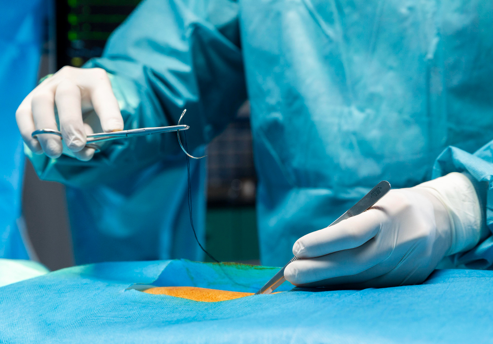
Atlanto Axial Surgery

Atlantoaxial surgery is a specialized procedure that focuses on addressing abnormalities and instability between the atlas (C1) and axis (C2) vertebrae in the upper cervical spine. This region is critical for providing stability and mobility to the head and neck. Surgery may be necessary to alleviate compression of the spinal cord or nerves, correct deformities, or stabilize the spine following trauma or degenerative changes.
Common Indications for Atlantoaxial Surgery
Atlantoaxial Instability
- Instability between the atlas and axis vertebrae, often due to trauma, congenital abnormalities, or inflammatory conditions like rheumatoid arthritis.
Atlantoaxial Dislocation
- Dislocation of the atlas relative to the axis, leading to compression of the spinal cord or nerve roots.
Atlantoaxial Subluxation
- Partial dislocation of the atlantoaxial joint, causing instability and potential neurological symptoms.
Congenital Abnormalities
- Conditions such as os odontoideum (an abnormality of the odontoid process) or atlas assimilation (fusion of the atlas with the occipital bone), which may require surgical correction to prevent neurological complications.
Procedure
Preoperative Evaluation
- Imaging Studies: MRI, CT scans, or dynamic X-rays to assess the extent of instability or compression and plan the surgical approach.
- Neurological Assessment: Evaluation of neurological symptoms and function to guide treatment decisions.
Surgical Techniques
- Posterior Fusion: The most common approach involves accessing the atlantoaxial joint from the back of the neck and fusing the vertebrae using bone grafts and screws or rods to stabilize the spine.
- Decompression: If there is compression of the spinal cord or nerves, decompression may be performed alongside fusion to relieve pressure.
- Transoral Approach: In select cases where there is anterior compression of the spinal cord, surgeons may access the atlantoaxial region through the mouth (transoral approach) to remove the offending structures and stabilize the spine.
Instrumentation
- Screws and Rods: Metal screws and rods are used to stabilize the vertebrae during fusion and promote bone healing.
- Bone Grafts: Bone graft material, either harvested from the patient’s own body or obtained from a bone bank, is used to facilitate fusion between the vertebrae.
Recovery
- Hospital Stay: Patients typically stay in the hospital for a few days to a week, depending on the complexity of the surgery and their postoperative recovery.
- Neck Immobilization: A cervical collar or brace may be worn to provide support and restrict motion during the initial healing phase.
- Physical Therapy: Rehabilitation exercises are initiated gradually to improve strength, mobility, and neck stability.
- Follow-Up Care: Regular follow-up appointments and imaging studies to monitor fusion progress and assess neurological function.
Benefits
- Stability: Restores stability to the atlantoaxial joint, reducing the risk of further dislocation or neurological complications.
- Pain Relief: Alleviates symptoms associated with spinal cord or nerve compression, such as neck pain, weakness, or sensory changes.
- Prevention of Neurological Damage: Helps prevent progressive neurological deficits by decompressing the spinal cord or nerve roots.
Risks and Considerations
- Infection: Risk of surgical site infection, which is mitigated by strict sterile techniques and postoperative antibiotic therapy.
- Hardware Failure: Potential for screws or rods to loosen or break, requiring revision surgery in rare cases.
- Neurological Complications: Risk of injury to the spinal cord or nerves during surgery, though this risk is minimized with careful surgical planning and intraoperative monitoring.
Atlantoaxial surgery is a highly specialized procedure that requires expertise in spinal anatomy and surgical techniques. When performed by experienced neurosurgeons, it can effectively address instability and compression in the upper cervical spine, providing patients with improved stability, pain relief, and preservation of neurological function.


